| 6 - Diatoms as siliceous and two-shelled cells | |
|
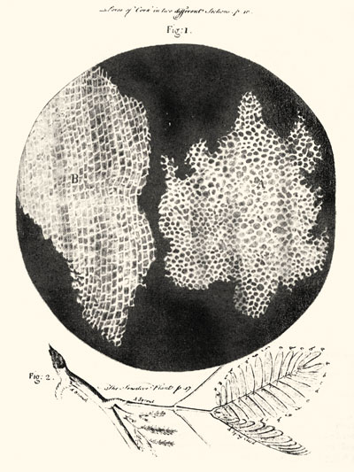
Cell. The word "cell" descends from the name for a room, especially a store-room, and hence usually a small room (Latin, cella, cellula). The Englishman Robert Hooke first used it in biology in his superbly illustrated Micrographia (1665) in describing his observations of the surface of a freshly cut piece of cork with the recently invented compound microscope (Figure 15):
For the next 150 years cells would remain not much more than these "little Boxes", chambers with solid walls detected primarily in plants or spaces enclosed by them, poorly understood structures with unknown functions and unknown importance. At the beginning of the 19th century, when Candolle wrote the Flore, the study of plant and animal structure remained almost entirely at the organ and tissue levels; it was still undeveloped at the cellular level. The concept of a cell as the fundamental unit of organic structure grew gradually out of the marriage of the microscopical observations of the 1820s and 1830s and the reductionist philosophical ideas of the German school of Nature Philosophy. This marriage was given scientific standing as the "cell theory" of Theodore Schleiden and Matthias Schwann in 1838-1839. And the concepts of "unicellular organisms" and of "all cells coming from pre-existing cells" were given their first argued presentation in the works of Karl Nägeli in the mid- to late-1840s. Thus, our contemporary concept of a cell did not precipitate until more than three decades after Flore française. Candolle uses "cellule(s)" in the Flore to mean simply a chamber -- as a chamber in a filament or in the stem of aquatic plant or in an ovule; his language at this time was the language of Linnaeus and Roth, not that of the next generation of botanists. |
Figure
15 (above). The cells in cork from Robert Hooke's Micrographia.
|
|
Figure
16. Friedrich Traugott Kützing (1809-1893). Image: Nordhausen am
Harz, http://www.nordhausen. |
Siliceous. Diatom cell walls are composed of silica, basically molecules of silicon and oxygen atoms polymerized in a highly regular, three-dimensional crystalline lattice. The domain of strong lattice regularity varies spatially in silica -- from that maintained over long distances, as in quartz, to that maintained only on a shorter scale, more locally, as in the amorphous silica of diatoms. Regardless of this microscale variation in geometry, the chemical analysis of silica, especially among mineralogists, was routine by 1800, and so Candolle could have subjected his diatomes to such an analysis, but he did not. Enter Friedrich Kützing (Figure 16) 25 years later, the apprentice pharmacist, Nordhausen school teacher, and indefatigable and prolific student of the algae. At the beginning of the 1830s the family of diatoms [Diatomeen or Bacillarien] included both diatoms and desmids. But Kützing as a result of his early microscopical researches had judged by 1833 a general difference between these two groups and separated them into families of their own:
He followed up on this suspicion of their "glassy" nature the following year. As a result of cleaning the calcium carbonate crust off a Chara, a large freshwater alga, with an acid so he could examine it, he noticed that the acid left unchanged the external parts of the diatoms which had been growing on it. After further experiments with heat and acids on other diatoms, he analzyed several different samples of them for silica and determined that the diatom "shell consists of pure silica." He was frustrated in getting his conclusions published, but they were finally noted in print by Christian Ehrenberg in 1835. Kützing emphasized this distinctive character of diatoms -- sufficient to separate them from desmids -- in the title of his important 1844 work,The silica-shelled bacillaria or diatoms [Die kieselschaligen Bacillarien oder Diatomeen]. |

|
|
Two-shelled. As importantly, also in his 1833 work, Kützing proposed for the first time that the external portion of diatoms was basically composed of two shells [thecae] (Figure 17):
|
|
Conclusions: That any organisms, much less diatoms, were understood to be unicellular (in any modern sense of the word) was not on firm ground until the 1840s. That diatom cell walls were either siliceous or bipartite was not shown before 1833-1834, almost 3 decades after the Flore. The knowledge Candolle needed to give the word diatome the meaning of the cutting-in-half of a "siliceous bipartite cell" was simply unavailable to him. |
|
| 5 - Lyngbye's interpretation |

Creative Commons Attribution-NonCommercial-NoDerivs 3.0 Unported License.